Treatment / Focus

Aneurysms can affect any artery but most commonly affect the aorta. The aorta runs from the heart through the chest and abdomen and gives off several very important branches supplying all internal organs, the spinal cord and the lower limbs.

An aortic aneurysm can occur in the chest (thoracic aortic aneurysm) or the abdomen (abdominal aortic aneurysm).
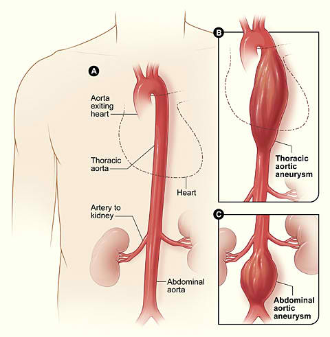
WHY IS AORTIC AN ANEURYSM DANGEROUS?

The normal aorta measures around 15 to 20 mm in diameter and has a strong and healthy arterial wall to sustain high pressure. Aortic aneurysms cause weakness in the wall of the aorta and increase the risk of aortic rupture.  The larger the aortic aneurysm the higher the chance of aorta bursting. When aneurysms rupture, blood suddenly escapes and this haemorrhage is life threatening. The consequences of aortic aneurysm rupture are disastrous and mortality is still between 50 to 70 %.

HOW CAN AN AORTIC ANEURYSM BE DIAGNOSED?

Aortic aneurysm is usually asymptomatic so patients are not aware of it. Men over 60 years are most frequently affected and those who have a positive family history may develop an aneurysm even earlier in life. An ultrasound scan is a reliable, pain free, quick and relatively inexpensive technique used to diagnose large aortic aneurysms. Ultrasound scans performed in a dedicated vascular laboratory is recommended for the above mentioned groups of patients.

HOW IS AN AORTIC ANEURYSM TREATED?

Every patient is unique and every aneurysm is different thus every case will be discussed with a consultant individually. In general, however, small aneurysms may be treated with medication and observation.

For larger or symptomatic aortic aneurysms several different treatment options exist:

Open aneurysm repair. Thoracic or abdominal aortic aneurysms can be repaired with a classical open surgical technique. This is a major procedure and the patient will be informed fully about all aspects. Also, in patients with other significant diseases there will be consultation with other specialists within our multidisciplinary team in order to deliver the best quality care.

Depending on the location of the aneurysm an incision will be placed over the tummy under general anaesthetic and the weakened aorta will be cut off and replaced with a synthetic tube graft. The patient stays in an Intensive Care Unit for the first several hours and then he/she will be transported back to their surgical department. The total hospital stay is usually one or two weeks.

Endovascular stent graft. This mini invasive, or keyhole, surgical procedure is mainly performed on patients who are medically not fit enough for open surgery. However, the patients may wish to have this procedure done even if they are fit for open surgery. The most important prerequisite is appropriate aortic anatomy. Everyone’s aorta will be thoroughly evaluated with a CAT scan or angiogram (dye test), measured and a custom made stent graft will be made for an individual patient if needed. The stent graft or endograft, will be introduced into the patient’s aorta using a catheter (a thin, flexible tube) inserted through a small incision into an artery in the groin. Under real-time x ray imaging the surgeon will guide the stent graft above and below the aneurysm and will exclude the weakened part of the patient’s aorta from the circulation.

General anaesthetic might still be required, but since the abdomen is not entered to accomplish the repair the patient usually leaves the hospital earlier then in the case of open aortic aneurysm repair. Postoperative follow up is necessary and CAT scan or ultrasound scans need to be done every year to make sure the stent graft is working well.

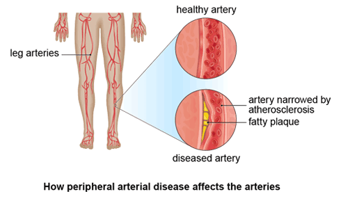
Peripheral arterial disease (PAD) is a condition in which arteries in the legs and less often elsewhere, is blocked by plaque. Due to a blockage in major arteries the circulation is impaired and leads to a variety of symptoms. Most, however, remain symptom free. When symptoms do occur, they include cramping pain or discomfort in the calf, thigh or buttocks when walking. Symptoms often cease very quickly when the patient stops walking. If the blockage affects several main stream arteries or multiple levels of the arterial tree then a patient’s feet and/or toes hurt continuously and the pain gets worse when the patient is lying down. Gangrene or non-healing ulcers can occur in cases of advanced arterial disease.

WHY IS PAD TREATED AND WHO IS AFFECTED MOST OFTEN?

Due to the lack of blood supply and therefore lack of oxygen, the tissue below the blockage is threatened. Where the foot or leg is affected the patient might loose his/her limb if the blockage is not repaired adequately or in time. Most often diabetes is associated with PAD. It is also the most common cause of lower limb amputation. Patients most likely to suffer from PAD are diabetics and cigarettes smokers.

HOW CAN PAD BE DIAGNOSED?

Every patient will be thoroughly assessed by a consultant and most of the time further testing will be required. A blood test and blood pressure measurement on both upper and lower limbs will be taken and either an ultrasound scan and/or CT scan will be performed. In cases where an intervention is planned an angiogram (dye test) will be performed by the vascular surgeon. All necessary information about the results of the tests will be thoroughly discussed with patient. The patient’s family doctor will be also informed with a letter regarding treatment options.

WHAT ARE THE TREATMENT OPTIONS AVAILABLE?

Every patient is unique and therefore a very individual approach will be taken to satisfy everyone’s needs. Not every arterial blockage needs to be treated surgically but those threatening limb viability or causing severe life-limiting limping should be treated by a vascular surgeon.

Open Revascularisation or Bypass. This classical surgical treatment is performed in situations where a blockage is not suitable for a mini invasive procedure (balloon angioplasty) or under other specific circumstances. Bypass is usually performed under a general anaesthetic or spinal anaesthesia and the patient’s native leg/thigh vein is used to bypass a long arterial blockage. A synthetic material also might be used. In the majority of cases a long incision is needed to harvest suitable vein graft and therefore recovery time in the hospital is about a week. Subsequent follow up scans are necessary during the first several years to make sure that the bypass is working well.

Endovascular treatment. This is the preferred treatment option for a short blockage or narrowing. It is a mini invasive option (keyhole surgery) with a short hospital stay. Every patient will be assessed thoroughly with an angiogram and a balloon angioplasty will be offered to every patient as a first treatment option if his/her arterial narrowing is found suitable for this treatment modality. Balloon angioplasty is a minimally invasive procedure in which the surgeon threads a balloon-tipped tube through the arteries until it reaches the one that is blocked. The surgeon inflates the balloon, which compresses the plaque in the artery and widens the vessel. Stenting is also minimally invasive and may be done at the same time as angioplasty. A stent is a small metal-mesh tube that a surgeon inserts to keep the artery open.

WHAT IS TOS?

The Thoracic Outlet represents a specific anatomic region between the collar bone, the first rib and muscles. Nerves and vessels pass through this narrow area and can get compressed there. Thoracic Outlet Syndrome  (TOS) develops in this case. The patient presents with various different symptoms including hand or arm swelling, skin colour changes, pins and needles in the forearm or hand, loss of sensation or even loss of strength in their hand on the affected side.

HOW CAN BE TOS DIAGNOSED?

Clinical assessment by a vascular surgeon is the best start. Further investigation is required in a majority of cases and includes ultrasound scan, CT scan and in some cases also angiogram (dye test). Neurological assessment can also be helpful.

HOW CAN TOS BE TREATED?

Initial treatment is almost always non-operative. The patient is examined by an experienced physiotherapist first and undergoes a trial of a range of tailor-made body exercises (physiotherapy). If symptoms do not improve over time surgical treatment is discussed with the patient. Based on underlying pathology found on a CT scan/angiogram a decompressing surgical procedure is planned and explained to the patient in detail including all possible alternatives. Typically the first rib is removed and nerves or vessels are relieved from being trapped in between muscles, tendons, bands and bones. The operation generally takes from one to two hours and the patient usually stays in hospital only for one or two days.

WHAT IS AN ARTERIOVENOUS FISTULA?

An arteriovenous fistula (AVF) is the connection of a vein and an artery. An AVF is surgically created, usually in the forearm and most often under local anesthesia, as a day-case. Patients go home the same day several hours following the operation. A more complex procedure should be expected if the patient does not have a suitable vein in the forearm. These more complex procedures are usually done under general anesthesia and require a one day hospital stay. An ultrasound vein assessment is necessary prior to every operation as the location and quality of the vein determines the type and site of surgical procedure.

WHY IS AN AVF CREATED?

The surgical creation of an AV Fistula provides a long-lasting site through which blood can be removed and returned during hemodialysis. Hemodialysis is a technique of ‘blood cleaning’ or ‘filtering’ through a machine in cases where a patient’s kidneys have stopped working. There is also a different type of dialysis called peritoneal dialysis. Only hemodialysis requires an AV Fistula or other vascular access. Both alternatives, however, need to be first discussed with a nephrologist (kidney specialist). The fistula should be created ideally several months before the patient starts their hemodialysis as AVF needs to mature.

WHAT TO DO FOLLOWING THE SURGERY?

Following creation of an AVF, every patient is seen by a specialised nurse and kidney specialist on a regular basis. Prior to the first hemodialysis every patient must also be seen by a vascular specialist to assess the quality of the AVF. Regular feedback from the dialysis nurse is desirable as any AVF can develop narrowing or even a blockage in the future and early and adequate intervention can save the fistula. Any AVF can also get infected or can develop an aneurysm. Those situations need to be assessed and treated by the vascular surgeon. Patients with an already existing AVF should be aware of the fact that long-term surveillance is needed to keep their AVF working well. Potential future interventions to keep the AVF well working are usually performed with minimally-invasive techniques as a day-case procedure (endovascular procedure).

Diabetes is associated with several long-term complications. Amongst the most devastating is lower limb amputation. The most frequent cause of amputation is the diabetic foot ulcer. Ulcers occur in about 10-25% of patients with diabetes and can lead to lower limb loss in up to 80 % of cases.

WHAT IS A DIABETIC FOOT ULCER?

Any skin breakdown or ulceration should heal in a timely fashion in healthy individuals. Diabetics tend to develop foot deformities possibly due to metabolic nerve injury (neuropathy). This can lead to an imbalance between certain groups of muscles controlling foot and toe movement. As a result of this imbalance diabetics tend to develop specific foot deformities such as hammer and claw toes and collapse of plantar arch. Diabetics also might not have good sensation in their soles (Sensory Neuropathy) and therefore can not feel pain. Also, skin tends to get dry and easily cracks if not adequately moisturised. Any foreign body in a patient’s shoe can cause a superficial wound that might not be felt by the patient due to a loss of protective sensation and can lead to a deep ulcer and limb loss later on. Most of the time, however, patients develop callus in the areas with high pressure due to underlying bone and joint deformities. This callus is abnormally hard skin which can lead to disruption of underlying tissue and can become infected. An ulcer then develops and this can affect underlying bones and joints. If not treated this situation can lead to limb loss and even to deterioration of the patient’s life functions.

Diabetics also tend to develop Peripheral Arterial Disease (PAD) and the distribution of the blockage and narrowing in their arteries has a very unique distribution. Unfortunately, most of the time small arteries below the knee level are affected, which makes treatment rather difficult. Lack of oxygen in such situations leads to non-healing ulcers or gangrene and without adequate treatment causes limb loss.

HOW IS DIABETIC FOOT AND ULCERS DIAGNOSED?

The important first step is taking a history of long-standing diabetes. Diabetic patients are usually seen by their podiatrist and also by a diabetologist and family doctor on regular basis. Their feet need to be seen either by the patient themself or their relatives, if they have visual disturbances. Every superficial wound should be seen by the patient’s family doctor and treated adequately. If healing is slow and non-responding to usual re-dressing it is time to refer the patient to a specialist. With a known history of diabetes previous ulcerations, foot deformities or even previous minor (toe) amputations, such referral should be obviously rather urgent. Podiatrists also are very important ‘gate keepers’ and their referral to a family physician and then to a specialist are highly appreciated. Diagnosis is based on ultrasound assessment of blood supply, measurement of the oxygen level in the foot skin, angiography and assessment of degree of neuropathy. In close cooperation with orthotics and a podiatrist a footprint is taken and plantar pressures are assessed. Foot X-ray and magnetic resonance imaging, or nuclear scan, might be necessary to diagnose deep tissue infection or osteomyelitis (bone infection).

HOW ARE DIABETIC FOOT ULCERS TREATED?

It is important to distinguish between different causes of diabetic foot ulcerations and then the treatment be tailored accordingly. For foot ulcers due to a lack of oxygen an adequate vascular or endovascular procedure is needed to improve the chances of healing (please see also PAD). Special skills are needed for such situations as diabetic’s arteries that need to be reconstructed are very stiff, small and fragile. Microsurgical skills are required for advanced foot-salvage procedures including soft tissue resurfacing. A surgical bypass or even a combination of bypass and free tissue transferred from distant part of the body can resurface even large foot defects and salvage the leg.

Neuropathic ulcers take, on average, more then 30 weeks to heal and their recurrence rate is high. In some situations where healing wasn’t successful and/or recurrence is unacceptable for a patient, a special reconstructive surgical procedure is possible to resurface the ulcerated area and correct underlying skeletal deformity. For such situations the patient is seen by other members of a multidisciplinary team. This includes orthopedic surgeons with special skills, podiatrists, orthotics and vascular surgeons who specialise in diabetic foot disorders. Every patient is thoroughly examined, the limb is assessed and then all reasonable therapeutic alternatives are discussed with the patient. Treatment is possible in a vast majority of situations and the outcome can be reliable, satisfactory and can significantly improve the patient’s quality of life. Postoperative preventive measures are crucial and can prevent development of further ulcers.

A large spectrum of venous disease exists and appears in various manifestations in patients of all ages. Varicose veins are superficial limb veins that have become stretched and swollen with blood. It is one of the most common vascular diseases in industrial countries. Multiple factors contribute to the development of varicose veins and include hereditary (family history) and environmental factors (occupation with prolonged standing, pregnancy etc). Other venous diseases include small reticular veins within the skin, deep vein thrombosis (DVT) and post thrombotic syndrome with one of the most devastating complications including skin ulcer. Also, pulmonary embolism might be associated with lower limb DVT.

WHAT DOES VENOUS INSUFFICIENCY CAUSE?

Most patients suffer from subjective symptoms like pain, soreness, burning or aching and sometimes even cramping pain and tiredness of the feet and legs. Swelling is often present. Over time, chronic non-treated venous insufficiency leads to cutaneous changes in terms of brown skin colouring or even soft tissue breakdown, which is known as ulceration.

HOW IS VENOUS INSUFFICIENCY DIAGNOSED?

Following a clinical appointment an ultrasound scan is requested and results are discussed with the patient. Further and more detailed assessments are needed in complex cases where previous surgical procedures have been performed or deep vein thrombosis has caused skin ulcerations and reconstruction of deep veins is planned.

HOW IS VENOUS INSUFFICIENCY TREATED?

Small skin veins can be treated for mainly cosmetic reasons by sclerotherapy on an outpatient basis. Varicose veins are usually treated with a surgical procedure and it is, most of the time, also an outpatient procedure. The goal of this operation is to remove insufficient (incompetent) veins that are causing symptoms, including those that are cosmetically unacceptable for the patient. The surgical procedure can be performed either under general anesthesia or local anesthesia. It consists essentially of one skin cut in the groin, or behind the patient’s knee and multiple small punctures. Through those small punctures incompetent veins are removed and the leg is then softly bandaged. Recovery time is about a week at home. Hospital stay is not required for the average, healthy patient.

The most advanced technology available involves using a laser or radiofrequency energy to seal larger faulty veins with no incision. This technique is available for most patients in our practice. Foam sclerotherapy and/or removal of the varicose veins can be done simultaneously or in stages, also under local anesthetic.

Deep vein reconstruction is sometimes necessary to improve venous circulation and heal long-lasting leg ulcers. Most commonly an endovascular venoplasty and stenting is used to repair obstructions in the deep veins. The procedure is very similar to one used for arterial occlusive disease (please see Peripheral Arterial Disease, balloon angioplasty). Specific surgical procedures are sometimes needed for patients with leg ulcers in cases where previous superficial vein surgery wasn’t successful. This procedure is mainly performed using minimally invasive techniques such as endoscopy and Subfascial Endoscopic Perforator Surgery.

In acute DVT situations where the large pelvic vein is affected, an intervention (thrombolysis) is also advised in order to ‘de-clot’ the vein, restore the vein’s flow, avoid a leg ulcer and chronic swelling associated with pain.

Pelvic pain, urinary urgency, vulvar varicose veins and many other symptoms can also be caused by enlarged vein in the pelvis. Obstruction or valve incompetence can be blamed and investigation usually involves CT scan and ultrasound. Treatment is efficient and key-hole based. For details please look at the Education section (questionnaire).

PRIMARY VARICOSE VEINS

Uncomplicated varicose veins can be treated at any point of time and treatment should be minimally invasive and cosmetically acceptable. The recommended treatment is endovenous laser ablation or radiofrequency ablation of the major subcutaneous veins (Great or Short Saphenous Veins) with or without additional phlebectomy or sclerotherapy (veins are either removed from the leg via small incision not larger then few millimeters or are injected with a special substance that makes them shrink). Everyone with varicose veins deserves assessment including ultrasound scan and based on the findings he or she can be offered adequate treatment.

Patients with more advanced CVD (significant leg swelling not improving with stockings, skin pigmentation or even skin ulcer) should be treated as soon as possible. Leg ulcers are very difficult to heal and if they do heal they tend to reoccur (in up to 70% of cases they reopen again).  The causes for these ulcers may be large primary varicose veins, previous deep vein thrombosis (DVT) or a vein compression (often between an artery and spine). For all patients with advanced CVD there is a realistic chance to improve their quality of life by a key-hole surgery (or mini-invasive surgical procedure under local anesthetic) with no overnight hospital stay. Results of this surgery have proved this is a very safe and reliable treatment.

ADVANCED CHRONIC VENOUS DISEASE

Patients with chronic swelling, skin pigmentation, redness or leg ulcers should seek a vascular specialist as their venous system is not working correctly.

Causes of chronic venous insufficiency are either “faulty valves” (incompetence or reflux) or a “blockage” in the deep veins (obstruction or compression). There are also often both obstruction and reflux found in many patients.

The concept of treatment of chronic venous insufficiency aims to:

  • 1. Reduce (eliminate) obstruction
  • 2. Reduce (eliminate) reflux (“faulty” valves in the veins)

Diagnosis is made by:

  • Ultrasound assessment (by expert technician)
  • CT Venogram/ MR Venogram – can give us more details about pelvic veins but up to 50% of blocked veins are underdiagnosed (blockage not seen) due to many issues (shape of the vein, non-contrast blood etc.)
  • Formal venogram (or angiogram) – has much better sensitivity rate of underdiagnosed blockages
  • IVUS (intravascular ultrasound) – the golden standard (unfortunately not funded and not always available)

The obstruction can be found in the venous system after DVT quite often. This is partially since in some segments of the venous system the vein after DVT often reopens incompletely despite adequate medical management (anticoagulation medication). Typical example is iliac (pelvic) or femoral (groin) veins. Obstruction increases venous pressure (Venous Hypertension) and is responsible for tissue damages.

In many people there is an asymptomatic (‘silent’) blockage waiting for a trigger like heart failure, inflammation, dehydration, etc. to become symptomatic – causing a non-thrombotic obstruction or acute DVT (a theory of a ‘permissive’ iliac vein lesion).

In certain complex situations where both reflux and obstruction play a role we treat one problem first. If the healing is satisfactory then no more intervention is required. If on the other hand, the healing is not as fast as we would have liked, another procedure is considered. It is mostly a combination of key-hole stenting (angioplasty) and phlebectomy (with or without laser surgery) that is usually opted for. In most of cases this is regarded as a day-procedure.

Redressing of the wounds in cooperation with a general practitioner or wound nurse is necessary for a certain period. Elastic compression stockings might not be necessary if the obstruction (and/or reflux) was successfully removed. There is a chance for patients who were wearing stockings to not to wear them afterwards at all. Anticoagulation medication (warfarin, dabigatran, etc.) in some situations is required and also aspirin (or Plavix) can be prescribed due to a previous DVT or stent placement, but in certain cases even those medications might be ceased, and the patient might not take any of them following a successful operation.

Cooperation with hematologists, dermatologists, wound nurses and general practitioners is necessary during and after the treatment of CVD. Patients are also encouraged to look for a second opinion.

Technology is advancing every year and there are a number of new devices available that may change a patient’s quality of life. If a previous physician’s intervention failed several years ago it does not mean that the next intervention will fail again. It might be the right time to discuss your leg problems with a different vascular specialist in Perth and revisit your options.

ACUTE DEEP VEIN THROMBOSIS (DVT) AND ITS CONSEQUENCES

Acute DVT can clot off any vein in the body. If the pelvic vein gets affected the full recovery is seen in only the minority of cases. Calf or thigh veins reopen more frequently by the body’s inner “clot dissolving” mechanism after several weeks. Therefore we advocate an intervention for pelvic vein thrombosis (Iliofemoral DVT) as soon as possible. This window of opportunity is 1-2 weeks and therefore patients have a time to seek adequate medical attention. General practitioner should put the patient on anticoagulation medication and refer them to a physician specialising in venous disease. Treatment method of choice is a pharmaco-mechanical thrombolysis, where a small catheter is placed into a vein and the clot is sprayed with a solution that allows it to dissolve. The clot is then sucked out clearing the vein. In most cases there will be an underlying blockage in the vein which might have triggered the DVT in the first place and this obstruction needs to be repaired by placing a stent in the vein. The stent is a flexible but strong (usually nitinol) spring that will keep the vein open. This procedure also can be done as an outpatient with no hospital stay.